To support hospitals and specialists around the world in meeting their safety standards requirements, the IAEA has produced a series of video tutorials on quality and dosimetry aspects of computed tomography. The new e-learning material will enhance the quality of diagnostic care for cancer, cardiovascular diseases, neurological disorders and other conditions for patients worldwide.
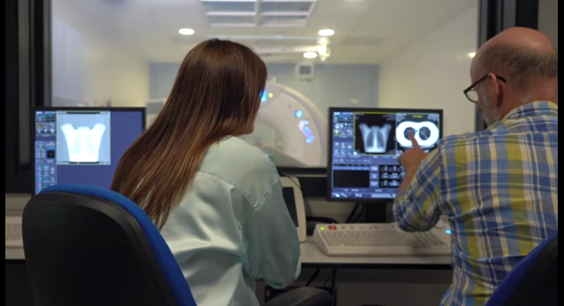
Computed tomography (CT) utilizes X rays and computer processing to visualize structures and soft tissues within the body. Through detailed, cross-sectional and three-dimensional volumetric images, CT scanners enable medical specialists to diagnose diseases, identify injuries, plan medical treatments, guide procedures and monitor disease progression as well as treatment effectiveness.
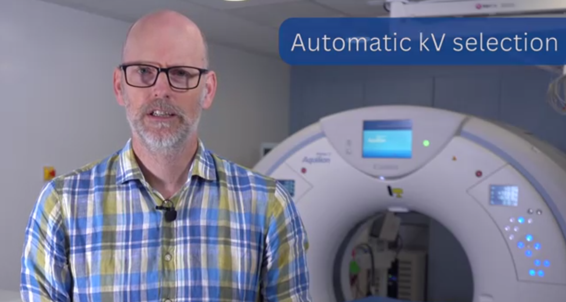
The target audience for the series is medical physicists, who play a key role in balancing imaging quality and radiation dose.
“The videos have been designed to help medical physicists get started with optimization of clinical imaging protocols. They emphasize the need to collaborate with clinical colleagues and provide an overview of key principles such as CT automatic exposure control, protocol management, and image analysis,” said David Platten, a clinical scientist at United Lincolnshire Hospitals NHS Trust in the United Kingdom and one of the global experts who contributed to the tutorials. “Implementing the principles described in the videos will enable centres to improve clinical practice through the optimization of CT image quality and patient radiation dose.”